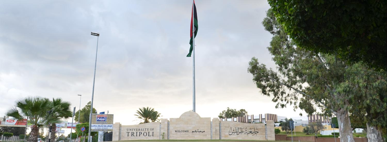
Department of Surgery & ٍٍReproductive

More ...

About Department of Surgery & ٍٍReproductive

Facts about Department of Surgery & ٍٍReproductive

We are proud of what we offer to the world and the community

14

Publications

10

Academic Staff

Who works at the Department of Surgery & ٍٍReproductive

Department of Surgery & ٍٍReproductive has more than 10 academic staff members

staff photo

Prof.Dr. lutfi Musa sasi BenAli

ا.د. لطفي بن علي هو احد اعضاء هيئة التدريس بقسم الجراحة والتناسليات بكلية الطب البيطري.انخرط بالسلك التدريسي بجامعة طرابلس كمعيد مند (1988) والان يشغل درحة استاد منذ 2015 وله العديد من المنشورات العلمية في مجال تخصصه

Publications

Some of publications in Department of Surgery & ٍٍReproductive

Patellar luxation in Hejazi goats

Background: Patellar luxation (PL) is a common orthopedic affection among farm and pet animals with mostly congenital (environmental and/or genetic) background. Aim: We report here the first observation of lateral PL in Hejazi goats bred in Libya. Methods: Five Hejazi goats aged between 4 months and 2 years with severe hind limb lameness were admitted to Al-Sorouh veterinary clinic in Tripoli during the period from 2016 to 2018. The goats were thoroughly examined clinically and radiographically. Two goats were surgically treated, and the other three cases were not because of either the cost limitation or expected poor prognosis. The surgical intervention involved femoral trochlear sulcoplasty, medial joint capsule imbrication, and tibial tuberosity transposition. Results: The clinical examination showed grade III–IV lateral PL. Radiologically, there were unilateral or bilateral, ventrocaudal, and dorsal PLs. Two cases were referred to surgical correction. One case almost restored the normal movement of stifle joint together with a good general status 1 year postsurgery. However, the surgical treatment was not effective in correcting the luxated patella in the second case. Conclusion: Lateral PL is common among orthopedic affections in Hejazi goats in Libya, and its surgical treatment provided a quite convenient approach. An association between inbreeding and the PL was suggested in those cases. Keywords: Clinical and radiological findings, Hejazi goat breed, Inbreeding, Patellar luxation, Surgical treatment.
Mohamed Hamrouni S. Abushhiwa, Abdulrhman Mohamed Salah Alrtib, Taher N. Elmeshreghi, Mouna Abdunnabi Abdunnabi Abdunnabi, Mansur Ennuri Moftah Shmela, Emad M R Bennour(3-2021)
Publisher's website

Normal biodistribution of canine leukocytes labeled in whole blood with technetium-99m stannous fluoride colloid and their ability to localize induced inflammatory foci

This study assessed the biodistribution of autologous leucocytes radiolabelled with technetium-99m stannous fluoride colloid (99mTcSnC) for detection of foci of induced inflammation in dogs. Venous blood was collected from seven healthy dogs and incubated with 99mTcSnC for 1 h at room temperature. Radiolabelled samples were injected intravenously (IV) and the dogs were scanned using a gamma camera. Another seven healthy dogs were injected intradermally with tumour necrosis factor α and then IV with 99mTcSnC radiolabelled autologous blood 3 h later before being scanned. The radiolabelled leucocytes localised to sites of inflammation by 30 min post-injection. IV injection of autologous leucocytes radiolabelled with 99mTcSnC appears to be a sensitive method for localisation of induced foci of inflammation in dogs.
Mohamed Hamrouni S. Abushhiwa, Nouria S.Salehi, Meir Lichtenstein, Peter M. Lording, Peter J. Finnin, Robert C. Whitton, Jennifer A. Charles, Bruce W. Parry (8-2010)
Publisher's website

First phalanx exostosis in traditional equestrian horses in Western Libya

Background: Orthopedic diseases involving the forelimb fetlock joint of horses other than those used in professional racing have not been widely reported in the literature. One of these problems is the exostosis on the proximal forelimb phalanx (P1), which has not been reported previously in Western Libya. Aim: The study aimed to investigate the prevalence of P1 exostosis in traditional equestrian horses (known locally as Sarj horses) of different breeds in Libya that participated in a special social event and described its clinical findings radiographic appearance. Methods: The current study involved 60 horses admitted to private veterinary clinics with varied fetlock orthopedic problems. The studied horses were aged between 6 and 11 years old and galloped on hard grounds. Clinical and radiographic examinations were carried out on all horses. Lateromedial radiographs for one or both forelimb fetlock joints were acquired for each horse. Results: Clinical examination revealed that 21 horses (35%) had hard non-painful swelling on the dorsal aspect of fetlock with joint stiffness during flexion. In 19 of them, the swelling was bilateral. No forelimb lameness was noticed. Radiographically, hard swelling was diagnosed as bone exostosis, with various sizes, on the proximo-dorsal aspect of P1. There was also a new bone formation on the disto-dorsal part of third metacarpal bone in two cases. Conclusion: This study has documented the first report on bone exostosis on the proximo-dorsal aspect of the forelimb proximal phalanx as a common problem in Sarj horses that galloped on hard grounds in Western Libya. Although this bone lesion did not cause lameness in all reported cases, further investigations are warranted to identify its histopathological nature, potential etiology, and proper treatment. Keywords: Exostosis, Fetlock, Forelimb proximal phalanx, Horse, Radiography.
Mohamed Hamrouni S. Abushhiwa, Taher N. Elmeshreghi, Abdulrhman Mohamed Salah Alrtib, Emad M R Bennour, Aiman Hussein Saleh Oheida(1-2022)
Publisher's website