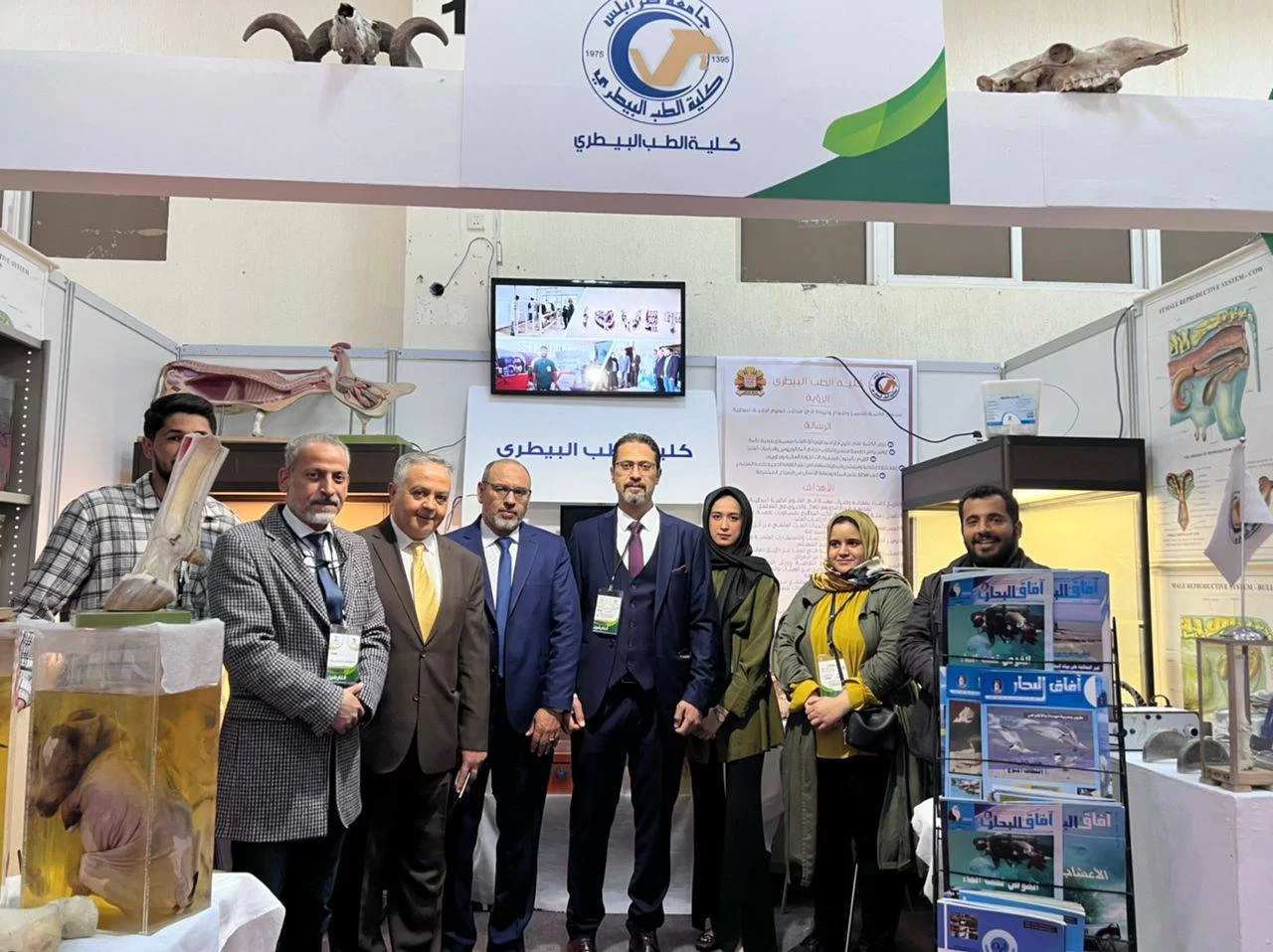
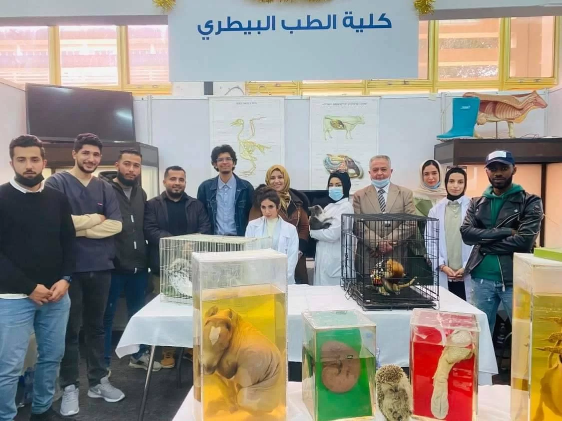
Faculty of Veterinary Medicine

More ...

About Faculty of Veterinary Medicine

The Faculty of Veterinary Medicine was established in 1975. It was the first Faculty of Veterinary Medicine in Libya. It is one of the citadels of science and knowledge at the University of Tripoli. This scientific institution works around the clock to meet the needs of the community of veterinarians and contributes to supporting the national economy. It values the care for animal health. It maintains increasing animal production, preserving human health and protecting the environment.

Facts about Faculty of Veterinary Medicine

We are proud of what we offer to the world and the community

194

Publications

86

Academic Staff

245

Students

23

Graduates

Programs

No Translation Found
Major No Translation Found

No Translation Found...

Details
Master of Poultry diseases
Major Veterinary medicine

This program is implemented through the study of academic courses, so that the number of units is not less than (24) and not more than (30) units of study over 3 semesters, in addition to the completion of a specialized scientific research thesis with (6) credits. The legal period required to obtain...

Details

Who works at the Faculty of Veterinary Medicine

Faculty of Veterinary Medicine has more than 86 academic staff members

staff photo

Dr. EHAB ِِAbdalla Tayeb Sharif

الأسم رباعي: إيهاب عبدالله الطيب الشريف مكان الميلاد: 1966 طرابلس – ليبيا الوضع الإجتماعي: متزوج المؤهـل العـلمي الحـالي:- درجة الدكتوراة البحثية. مجـــال التخصص في: مجال علوم البحار وأحياء البيئة البحرية. سنة الحصول عليه: 2013 – 2017 من معهد البيئة البحرية الساحلية (IAMC-CNR) – إيطاليا. - دبلوم متقدم في مجال البيئة المائية سنة 2011 من جامعة كولونيا – ألمانيا. - دبلوم العلاقات الدبلوماسية و السياسية و المنازعات ذات الطابع الدولي. المـؤهل الجـامعي : بكالوريس طب بيطري – كلية الطب البيطري - جامعة طرابلس – ليبيا سنة الحصول عليه: 1990-/ 1991 جامعة طرابلس. المجالات و الإهتمامات:  أمراض الأسماك , البيئة والتلوث البحري .  مراقبة و جودة المنتجات السمكية.  الهندسة الوراثية والتقنيات الحيوية البحرية.  الزراعات المائية.  المتاحف البحرية الخبرة, التعين والتاريخ الوظيفي: 1. العمل كطبيب بيطري بمستوصف عين زارة البيطري من سنة 1990 - 1993. 2. العمل كطبيب بيطري بمحطات أبحاث الأبقار والأغنام بكلية الزراعة / جامعة طرابلس من 1993 – 1995. 3. باحث مختص أمراض الأسماك والأحياء الدقيقة بمركز بحوث الأحياء البحرية من 1995 حتى 2019. 4. مدقق بالمركز الوطني لضمان جودة وإعتماد المؤسسات التعليمية. 5. عضو(سابق) باللجنة الفنية المتخصصة في مجال مواصفات وجودة السلع والمنتجات البيطرية التابع للمركز الوطني للمواصفات والمعايير القياسية. 6. عضو اللجنة الدائمة لمنح المؤسسات تراخيص تصدير المنتجات السمكية الليبية وضبط الجودة للأسماك ومنتجات الصيد البحري المصدرة بمركز تنمية الصادرات الخاصة. 7. عضو باللجنة العليا لتسجيل الأدوية والمستحضرات الطبية البيطرية. 8. عضو لجنة إدارة الهيئة العامة للثروة البحرية.

Publications

Some of publications in Faculty of Veterinary Medicine

Molecular Identification, Prevalence and Antimicrobial Susceptibility Profile of Cronobacter spp. Cultivated on a Chromogenic Medium in Libya

Background: Cronobacter sakazakii is associated with illness in infants from contaminated powdered infant formula (PIF) and it is frequently recovered from PIF factory environment. Limited information is available on contamination of other food such as dairy and meat products in Libya. Methods and findings: A total of 261 samples of milk, dairy products and coarse ground meat products were collected from different localities in Libya. Samples were examined for Cronobacter spp. with an adapted ISO /DTS 22964 cultural protocol using HiChrome™ Enterobacter sakazakii modified agar coupled with 16S rDNA partial sequencing to identify the organism. The identified isolates were biochemically characterized and tested for their ability to produce yellow pigment. Out of the 261 analyzed samples, only two beef burgers, one fermented milk “Laban”, one she-camel’s milk, two raw cow’s milk, two cereal baby food, one Maassora cheese and one ready to feed baby milk were contaminated with Cronobacter spp. at a total rate of 3.8%. Accuracy of HiChrome Ent. sakazakii modified agar reach 100% as all of blue-green presumptive colonies were confirmed Cronobacter spp. while other colorless, greenish or with blue center colonies which competed growth with Cronobacter spp. were predominantly Escherichia coli followed by Klebsiella spp. and to less extent Pseudomonas luteola, Citrobacter freundii and Acinetobacter baumanii. Moreover, the isolated strains of Cronobacter were resistant to Amoxicillin, Erythromycin, Vancomycin and Streptomycin, and sensitive to Doxycycline, Enrofloxacin and Gentamycin. Conclusion: This study documents for the first time the occurrence of Cronobacter spp. in beef burger, raw cow’s milk, fermented milk “Laban”, she-camel’s milk, Maassora cheese, cereal baby food and ready to feed baby milk sold in Libya, by using conventional methods, biochemical tests and molecular techniques.
Ibrahim Eldaghayes(12-2017)
Publisher's website

Effect of low and high doses of Salmonella enteritidis PT4 on experimentally infected chicks

Chicks (1-d-old, three groups, each containing 50 chicks) were inoculated with 2×10² and 2×10⁸ CFU ofSalmonella enteritidis; the third group were kept as uninoculated control. Five birds from each group were euthanized at intervals from 6 h to 4 weeks post-inoculation (pi). In the lowdose groupS. enteritidis was isolated from 60% cecal samples at 18 h pi. and from 20% of livers at 3 d pi. Individual variation in the frequency ofS. enteritidis recovery was observed in this group. The clearance of salmonella from the organs was faster in the low-dose group, and salmonella was not isolated from the liver and cecum at 21 and at 27 d, pi, respectively. However, in the high-dose group,S. enteritidis was isolated from all ceca and 80% of liver 6 h pi, and salmonella was detected in the cecum and liver throughout the experiment. Serous typhlitis and unabsorbed yolk sac were the most prevalent lesions in both groups. Granulomatous nodules in the cecum were found occasionally in some cases in both inoculated groups, which can play a role as reservoirs in carrier chicks.
A. A. Asheg(10-2001)
Publisher's website

Evaluation of Thoracic Limb Loads, Elbow Movement, and Morphology in Dogs Before and After Arthroscopic Management of Unilateral Medial Coronoid Process Disease

Objective To (1) evaluate thoracic limb loads and symmetry, and elbow function and morphology, before and after arthroscopic treatment of unilateral medial coronoid process disease (MCPD), and (2) determine if functional variables correlate with morphologic findings.Study DesignProspective case series.AnimalsDogs (n = 14) with thoracic limb lameness.Methods Dogs were included when unilateral MCPD was confirmed as the cause of lameness. Kinetic analysis of both thoracic limbs, along with kinematic analysis and goniometry of both elbows were carried out before, and 60, 120, and 180 days after partial coronoidectomy by arthroscopy. Radiography and computed tomography of both elbows were performed before and 180 days after arthroscopy.ResultsA nonsignificant (P = .11) increase in the peak vertical loads (PFz), and a significant (P = .022) increase in the vertical impulse (iFz) applied by the affected limb were seen. Symmetry indices improved, with significant differences between sessions (PFz: P = .019; iFz: P = .003). Kinematic variables showed no significant differences, between sessions or when comparing both elbows within sessions. Goniometry revealed no significant differences between sessions, but some significant differences were identified when comparing both elbows within sessions. Osteophytosis and degree of lameness showed no correlation, before (rs = −0.077; P = .79) or after arthroscopy (rs = 0.27; P = .35).Conclusions Kinetic variables improved after arthroscopy, without full restoration of function. Kinematic variables did not change significantly. Osteoarthritis and goniometric measurements in the affected joint worsened. Functional variables did not correlate with morphologic findings. arabic 23 English 137
Jalal Mohamed Abdelhadi(7-2004)
Publisher's website

Journals

Journals published by Faculty of Veterinary Medicine

Faculty of Veterinary Medicine Video Channel

Watch some videos about the Faculty of Veterinary Medicine

See more

Faculty of Veterinary Medicine in photos

Faculty of Veterinary Medicine Albums